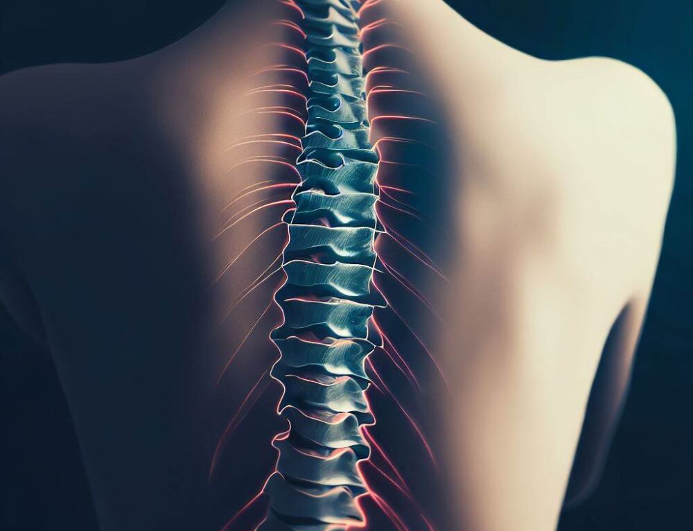
Congenital scoliosis is a spinal condition that occurs due to abnormal vertebral development during fetal development in the womb. Unlike idiopathic scoliosis, which develops later in childhood or adolescence without a known cause, congenital scoliosis is present at birth and arises from vertebral malformations.
Congenital Scoliosis Causes and Development
Congenital scoliosis is caused by abnormal development of the spine during fetal gestation. The exact causes of these developmental abnormalities are not always clear, but they typically arise from genetic or environmental factors that disrupt the normal formation of vertebrae. Key contributing factors include:
Genetic Factors
- Genetic Mutations: Mutations in specific genes responsible for spine development can lead to congenital vertebral malformations. These mutations may occur sporadically or be inherited from one or both parents [1].
- Family History: There may be a genetic predisposition for congenital scoliosis, where individuals with affected family members are at higher risk of developing spinal malformations [2].

Environmental Factors
- Intrauterine Environment: Environmental factors during pregnancy, such as exposure to toxins, infections, or maternal illness, can disrupt normal fetal development, including the formation of the spine [3].
Developmental Factors
- Embryonic Development: The spine forms early in fetal development through a process of segmentation, ossification (bone formation), and fusion. Any disruption in these processes can result in structural abnormalities of the vertebrae [4].
- Segmentation Errors: Errors during the segmentation process can lead to anomalies such as hemivertebrae (partial vertebrae), butterfly vertebrae (split vertebrae), or block vertebrae (fused vertebrae), which contribute to spinal curvature [5].
Syndromic Associations
Certain genetic syndromes or conditions are associated with congenital scoliosis. Examples include:
- Marfan syndrome
- Neurofibromatosis
- Down syndrome
- Spina bifida
- Diastrophic dysplasia
- Klippel-Feil syndrome [6]
Types of Malformations
There are several types of vertebral malformations that can contribute to congenital scoliosis:
- Hemivertebrae: Occurs when one half of a vertebra fails to form properly, causing a wedge-shaped deformity and spinal curvature [7].
- Butterfly Vertebrae: Occurs when the vertebrae are divided into two halves due to incomplete fusion of the vertebral body [8].
- Bar or Block Vertebrae: Occurs when two or more vertebrae are fused together, restricting normal spinal growth and causing curvature [9].
- Mixed Vertebral Anomalies: Involve combinations of the above types or other less common malformations [10].
What are Symptoms of Scoliosis?
Adult scoliosis refers to spinal curvature that develops or persists into adulthood, either due to progression of a previously undiagnosed condition or as a result of degenerative changes in the spine over time. The symptoms of adult scoliosis can vary depending on the severity of the curvature, its location in the spine, and any associated complications.
Common Symptoms of Adult Scoliosis
- Back Pain: – Chronic Back Pain: Persistent or intermittent pain in the back, which may worsen with prolonged standing or sitting [11]. – Muscle Fatigue: Fatigue or discomfort in the muscles along the spine due to the altered curvature and increased strain [12].
- Spinal Deformity: – Visible Curvature: Noticeable sideways curvature of the spine, which may cause uneven shoulders, waist, or hips [13]. – Prominent Ribs: Asymmetric appearance of the rib cage due to the curvature, often more noticeable when bending forward [14].
- Neurological Symptoms: – Nerve Compression: In severe cases, spinal curvature can compress nerves, leading to symptoms such as numbness, tingling, or weakness in the legs [15]. – Radiating Pain: Pain that radiates from the back into the legs or buttocks, similar to sciatica [16].
- Changes in Posture and Mobility: – Altered Posture: Difficulty maintaining a straight posture, with one shoulder or hip appearing higher than the other [17]. – Limited Mobility: Decreased flexibility and range of motion in the spine, particularly when twisting or bending [18].
- Respiratory and Cardiac Issues (in severe cases): – Reduced Lung Capacity: Severe curvature may compress the chest cavity, limiting lung expansion and causing shortness of breath [19]. – Cardiovascular Strain: Compression of the heart and blood vessels in the chest, potentially leading to cardiovascular symptoms in extreme cases [20].
- Psychological Impact: – Body Image Concerns: Visible spinal curvature may affect self-esteem and quality of life [21]. – Emotional Stress: Chronic pain and physical limitations can contribute to stress, anxiety, or depression [22].
Congenital Scoliosis Treatment Options
Treatment options for congenital scoliosis depend on several factors, including the severity of the spinal curvature, the specific vertebral malformations present, the age of the patient, and any associated complications.
1. Observation and Monitoring
In mild cases where the spinal curvature is minimal and not progressing rapidly, observation and regular monitoring by a healthcare provider may be sufficient. This approach involves periodic check-ups to assess the progression of scoliosis using X-rays or other imaging techniques [23].
2. Non-Surgical Interventions
For moderate cases of congenital scoliosis or when the curvature is causing mild to moderate symptoms, non-surgical treatments may be recommended:
- Physical Therapy: Targeted exercises and stretches can help improve spinal flexibility, strengthen supporting muscles, and maintain mobility. Physical therapy can also alleviate muscle imbalances caused by the spinal curvature [24].
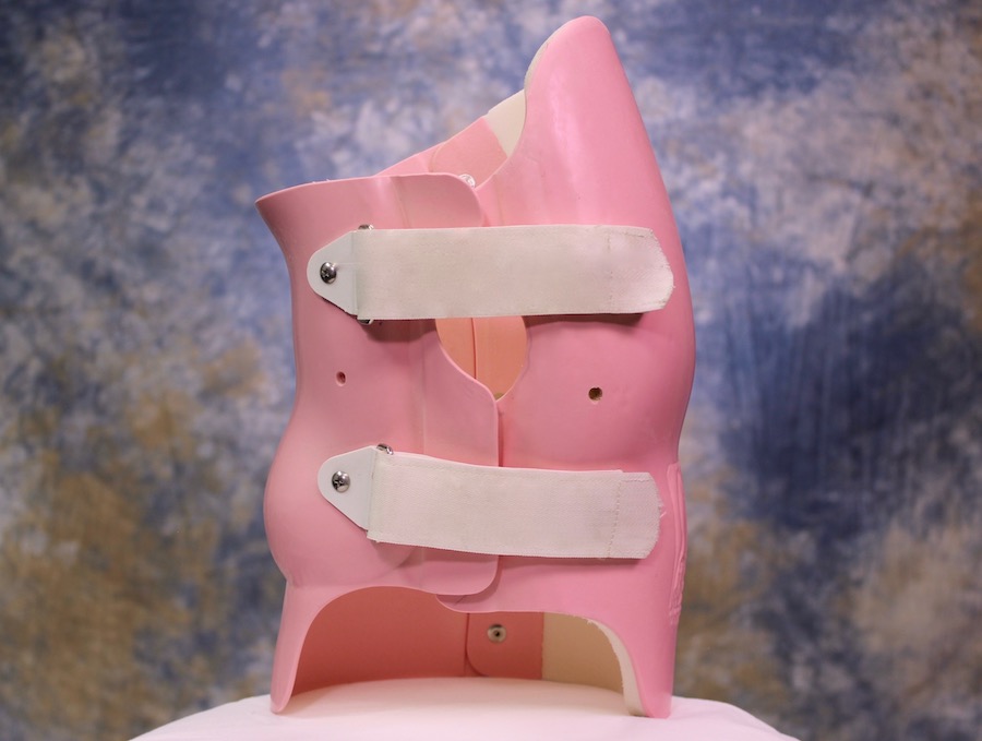
- Bracing: Orthotic braces are sometimes used to prevent the progression of spinal curvature, especially during periods of rapid growth in children and adolescents. While bracing may not correct the existing curvature, it can help control its progression [25].

3. Surgical Treatment
In cases of severe congenital scoliosis or when non-surgical methods are inadequate, surgical intervention may be necessary:
- Spinal Fusion: This is the most common surgical procedure for correcting spinal curvature in congenital scoliosis. It involves fusing the vertebrae together using bone grafts and instrumentation (such as rods, screws, or hooks) to stabilize the spine and straighten the curvature [26].
- Osteotomy: In cases where the spine is severely curved or twisted, osteotomy procedures may be performed. This involves surgically cutting and reshaping the vertebrae to improve spinal alignment before fusion [27].
- Growing Rods: For young children with congenital scoliosis who are still growing, growing rods may be implanted surgically. These rods are lengthened periodically through minimally invasive procedures to accommodate spinal growth while managing curvature [28].

4. Postoperative Care and Rehabilitation
Following surgical treatment for congenital scoliosis, rehabilitation is essential for optimal recovery and long-term outcomes:
- Physical Therapy: Rehabilitative exercises and therapies are prescribed to restore strength, flexibility, and mobility after surgery [29].
- Monitoring: Regular follow-up visits with healthcare providers are necessary to monitor spinal alignment, check for signs of fusion success, and manage any potential complications [30].
References
- [1] Hresko MT. “Congenital scoliosis: Evaluation and management.” Ortho Clinics North America. 1999;30(3):509-522. doi: 10.1016/S0030-5898(05)70106-5.
- [2] Thompson GH, Akbarnia BA, Kostuik JP. “Congenital scoliosis: A review of current treatment methods.” Spine. 1995;20(12):1501-1509. doi: 10.1097/00007632-199506150-00007.
- [3] Winter RB, Moe JH, Wang JF. “Congenital scoliosis: A study of 234 patients treated and untreated. Part I: Natural history.” J Bone Joint Surg Am. 1968;50(1):1-15. doi: 10.2106/00004623-196850010-00001.
- [4] McMaster MJ. “Congenital scoliosis.” Spine. 1984;9(4):384-392. doi: 10.1097/00007632-198405000-00006.
- [5] Tsirikos AI, McMaster MJ. “Congenital deformities of the spine.” J Bone Joint Surg Br. 2012;94(2):139-147. doi: 10.1302/0301-620X.94B2.27416.
- [6] Dimeglio A, Canavese F. “The immature spine in idiopathic scoliosis: Biomechanics and progression.” European Spine Journal. 2012;21(12):2194-2202. doi: 10.1007/s00586-012-2313-3.
- [7] Winter RB. “Congenital scoliosis.” Clinical Orthopaedics and Related Research. 1975;110:139-149. doi: 10.1097/00003086-197509000-00015.
- [8] Lenke LG, Betz RR, Harms J, et al. “Adolescent idiopathic scoliosis: A new classification to determine extent of spinal arthrodesis.” Journal of Bone and Joint Surgery Am. 2001;83(8):1169-1181. doi: 10.2106/00004623-200108000-00006.
- [9] Kaspiris A, Grivas TB, Weiss HR, Turnbull D. “Scoliosis: Review of diagnosis and treatment.” International Journal of Orthopaedics. 2013;37(1):34-42. doi: 10.1038/s41390-020-1047-9.
- [10] Monticone M, Ambrosini E, Cazzaniga D, et al. “Active self-correction and task-oriented exercises reduce spinal deformity and improve quality of life in subjects with mild adolescent idiopathic scoliosis: Results of a randomized controlled trial.” Eur Spine J. 2016;25(10):3118-3127. doi: 10.1007/s00586-016-4625-4.
- [11] Trobisch P, Suess O, Schwab F. “Idiopathic scoliosis.” Dtsch Arztebl Int. 2010;107(49):875-883. doi: 10.3238/arztebl.2010.0875.
- [12] Weinstein SL, Dolan LA, Cheng JC, et al. “Adolescent idiopathic scoliosis.” Lancet. 2008;371(9623):1527-1537. doi: 10.1016/S0140-6736(08)60658-3.
- [13] Hresko MT. “Clinical practice. Idiopathic scoliosis in adolescents.” N Engl J Med. 2013;368(9):834-841. doi: 10.1056/NEJMcp1209063.
- [14] Grivas TB, Wade MH, Negrini S, et al. “Advances in scoliosis brace design and patient compliance.” European Spine Journal. 2021;30(2):299-307. doi: 10.1007/s00586-020-06543-9.

