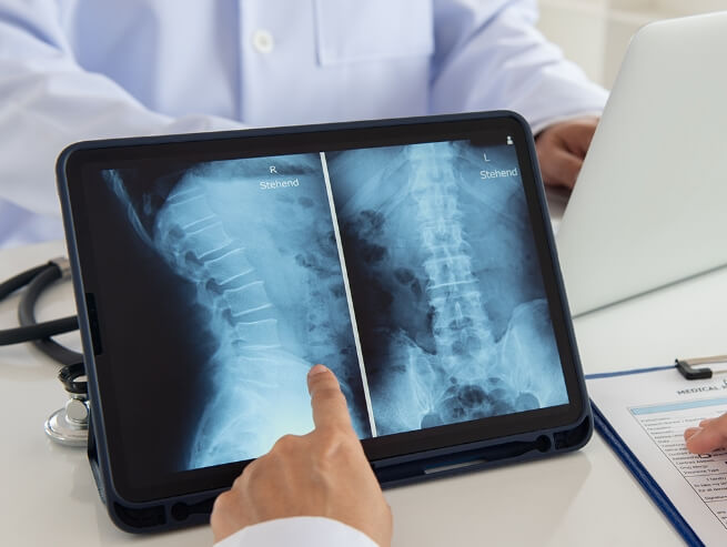
Verstehen Whole Spine X Ray
The utilization of radiation to create intricate images of the spinal bones characterizes a spinal X-ray. This diagnostic procedure plays a crucial role in helping physicians identify the root causes of back or neck pain experienced by patients.
The process involves a technician employing a machine that directs X-ray beams through the body, capturing a black-and-white image on specialized film or computer software. The resulting image distinguishes bones and other dense tissues, appearing white, from softer tissues such as fat or muscle, which manifest in varying shades of gray.
Why do You Need Whole Spine X Ray?
The X-ray will help you understand the reason of pain in your back or neck. Pain occurs due to a range of reasons such as injuries like a fracture, a break, infection, tumors or any other condition. It may also be ordered if bones have been reset after being broken to see if the bones are in perfect alignment or not.
X-rays are quick and painless procedures. They help the doctor in identifying abnormalities in any part of the spine. The imaging helps the doctors in drawing up appropriate treatment plans. They may also suggest lifestyle changes and set you on the path to recovery, relieving you from constant muscle pain.
The spine, composed of 33 small bones known as vertebrae, is divided into distinct sections:
- Cervical spine (seven vertebrae in the neck)
- Thoracic spine (12 vertebrae in the chest or trunk area)
- Lumbar spine (five vertebrae in the lower back)
- Sacral area (five small, fused vertebrae at the base of the spine)
- Coccyx (four coccygeal vertebrae fuse to form the tailbone)
Diverse Conditions Diagnosed by Spinal X-Rays
A spinal X-ray serves as a diagnostic tool for various conditions, including:
- Broken bones
- Arthritis
- Spinal disk problems
- Tumors
- Osteoporosis
- Abnormal spinal curves
- Infections
- Congenital spinal issues
While X-rays excel in pinpointing fractures and skeletal defects, they also aid in identifying connective tissue problems. Typically, these imaging studies confirm symptoms and examination findings to pinpoint the source of pain, particularly in cases of direct trauma to the back, back pain accompanied by fever, or weakness and numbness in the limbs.
Navigating the Landscape of Whole Spine X Ray Risks
While X-rays are generally safe, concerns about radiation-induced cellular changes leading to cancer exist. Nevertheless, the minimal radiation used in spinal X-rays mitigates this risk. Maintaining a record of prior X-rays and addressing long-term exposure concerns with a healthcare professional is advisable. It is essential to note that unborn babies are more susceptible to radiation, warranting disclosure of pregnancy to explore alternative imaging methods.
Preparing for a Whole Spine X Ray
Before undergoing a spinal X-ray, informing the healthcare provider about pregnancy, insulin pump usage, and recent X-rays, such as barium X-rays, is crucial. Additionally, removing metal items like jewelry, hairpins, eyeglasses, and hearing aids is necessary, as they may interfere with the imaging process.
The Whole Spine X Ray Procedure
During the examination, patients lie on a specialized table beneath an overhead X-ray machine. A trained technician positions the patient to ensure the section of the spine under examination aligns with the machine. A lead apron may be used to shield other body parts from radiation.
As the X-ray machine activates, patients are required to remain still and hold their breath momentarily. The process, lasting a few seconds, is painless, though accompanying clicking or buzzing sounds may be heard. Depending on the case, patients may stand for certain images or perform specific movements.
Upon completion, the technician processes the images, and patients may be asked to wait briefly for clarity confirmation.

Decoding Whole Spine X Ray Results
Following the examination, a healthcare professional, often a radiologist, reviews the images to analyze the spinal condition. Subsequently, patients engage in a discussion with their doctor, who provides insights into the findings and outlines the subsequent steps.
How whole spine x ray Works
Spinal X-rays operate based on the principles of high-frequency energy waves known as X-rays. These waves possess the ability to permeate through the body, interacting with target tissues in a process where absorption, reflection, or transmission occurs. As X-rays traverse the body, they can generate two-dimensional black-and-white images of the structures they pass through. These images are captured on a photographic film positioned on the opposite side of the body.
Specifically tailored to scrutinize the three primary segments of the spine—the cervical spine (neck), thoracic spine (mid-back), and lumbar spine (lower back)—as well as the pelvis, including the sacroiliac joint, spinal X-rays contribute to comprehensive diagnostic assessments.
X-ray Imaging of Spinal Bones Dense tissues characterized by a high calcium content, such as bones, exhibit reduced X-ray absorption, resulting in a white-colored representation on the photographic film. Bones emerge as the most distinct and clearly visible structures in X-ray images. The architecture, form, and alignment of spinal vertebrae and facet joints can be precisely documented through spinal X-rays.
X-ray Imaging of Spinal Soft Tissues Soft tissues permit a greater passage of X-rays, producing a dark impression on the photographic plate. Muscles, ligaments, and tendons, often implicated in back and neck pain, do not exhibit optimal visibility in conventional X-rays. Similarly, neural tissues, including the spinal cord and spinal nerves, responsible for potential back or neck discomfort, are typically inadequately defined in conventional X-rays. However, tumors, as masses of tissue, can be discerned in X-ray images due to their bulk and positioning.
Vorausschauende Planung‘s Advanced Technology in Whole Spine X Ray
As a leader in innovative medical technologies, Forethought integrates cutting-edge advancements in spinal X-ray procedures, ensuring accurate diagnostics and enhanced patient care. With a commitment to pushing the boundaries of medical imaging, Forethought contributes to the evolution of spinal healthcare.
Referenzen
- [1] Brenner DJ, Hall EJ. “Computed tomography—an increasing source of radiation exposure.” N Engl J Med. 2007;357(22):2277-2284. doi: 10.1056/NEJMra072149.
- [2] Modic MT, Ross JS. “Lumbar degenerative disk disease.” Radiology. 2007;245(1):43-61. doi: 10.1148/radiol.2451051359.
- [3] Ulbrich EJ, Schraner C, Boesch C, et al. “The lumbar spine: normal anatomy, variants, and pitfalls in MRI interpretation.” Eur J Radiol. 2010;74(2):149-159. doi: 10.1016/j.ejrad.2009.04.059.
- [4] Jensen MC, Brant-Zawadzki MN, Obuchowski N, et al. “Magnetic resonance imaging of the lumbar spine in people without back pain.” N Engl J Med. 1994;331(2):69-73. doi: 10.1056/NEJM199407143310201.
- [5] Weiser DA, Coley BD, Karmazyn B, et al. “MRI and Radiation-Free Imaging in Pediatric Radiology: Techniques and Applications.” Pediatric Radiology. 2021;51(3):354-367. doi: 10.1007/s00247-020-04858-9.
- [6] Smith JS, Shaffrey CI, Berven S, et al. “Spine surgery for pediatric and adult spinal deformity: an overview of the challenges and solutions.” J Pediatr Orthop. 2012;32(6):736-747. doi: 10.1097/BPO.0b013e31825bca0d.
- [7] Parent S, Newton PO, Wenger DR, et al. “Effectiveness and patient experience of radiation-free imaging modalities in monitoring scoliosis in adolescents.” J Pediatr Orthop. 2019;39(5):305-311. doi: 10.1097/BPO.0000000000001265.
- [8] Glassman SD, Berven S, Kostuik J, et al. “Scoliosis Research Society Instrument Validation Study: A Multicenter Assessment of Surgical Outcomes in Idiopathic Scoliosis.” Spine. 2005;30(6):699-702. doi: 10.1097/01.brs.0000157447.56975.3e.
- [9] Godzik J, Sielatycki JA, Chen H, et al. “Utility of radiation-free imaging in neurosurgical practice.” Journal of Neurosurgery. 2020;132(2):535-541. doi: 10.3171/2018.10.JNS182285.
- [10] Kalichman L, Cole R, Kim DH, et al. “Spinal stenosis prevalence and association with symptoms: the Framingham Study.” Spine J. 2009;9(7):545-550. doi: 10.1016/j.spinee.2009.03.005.
- [11] McCollough CH, Bushberg JT, Fletcher JG, et al. “Answers to Common Questions About the Use and Safety of CT Scans.” Mayo Clinic Proceedings. 2015;90(10):1380-1392. doi: 10.1016/j.mayocp.2015.07.011.
- [12] Weiser DA, Coley BD, Karmazyn B, et al. “MRI and Radiation-Free Imaging in Pediatric Radiology: Techniques and Applications.” Pediatric Radiology. 2021;51(3):354-367. doi: 10.1007/s00247-020-04858-9.
- [13] Tins BJ, Cassar-Pullicino VN, Lalam R, et al. “Spinal infection: the role of imaging.” Br J Radiol. 2014;87(1037):20130418. doi: 10.1259/bjr.20130418.
- [14] Gupta MC. “Neuromuscular scoliosis.” Journal of Orthopaedic Surgery. 2015;23(3):414-418. doi: 10.1177/230949901502300323.
- [15] Hricak H, Amparo EG, Crooks LE, et al. “MR imaging of the female pelvis: initial experience.” Radiology. 1983;148(1):159-165. doi: 10.1148/radiology.148.1.6856837.

