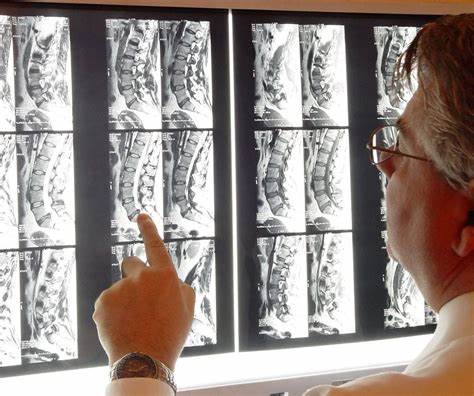
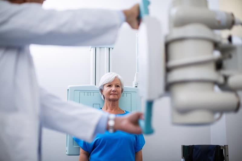
Scoliosis is a medical condition characterized by an abnormal curvature of the spine. It affects approximately 2-3% of the population, with the majority of cases occurring in adolescents. X-rays are commonly used to assess the severity and progression of scoliosis, providing valuable information for diagnosis and treatment planning. However, the accuracy of these assessments heavily relies on proper positioning during the X-ray procedure. This article will explore the importance of X-ray positioning in scoliosis assessment, discuss common X-ray positions, examine the advantages and disadvantages of supine and standing positioning techniques, identify factors affecting X-ray positioning accuracy, highlight best practices for scoliosis X-ray positioning, and emphasize the role of radiologists in ensuring proper positioning.

Skoliose verstehen
Before delving into the intricacies of X-ray positioning, it is essential to have a basic understanding of scoliosis. Scoliosis is a condition that causes the spine to curve sideways, resulting in an “S” or “C” shape. It can be classified as either structural or nonstructural, with the former involving a fixed curvature and the latter being reversible. Scoliosis can cause various symptoms, including back pain, uneven shoulders or hips, and limited mobility. X-rays play a crucial role in diagnosing and monitoring scoliosis, allowing healthcare professionals to accurately measure the degree of curvature and assess its progression over time.

Importance of X-Ray Positioning
Accurate assessment of scoliosis requires precise measurements of the spinal curvature. X-ray positioning plays a vital role in achieving this accuracy. Proper positioning ensures that the spine is adequately aligned and visible on the X-ray image, allowing for accurate measurements of the curvature angles. Incorrect positioning can lead to distorted images, making it challenging to assess the severity of scoliosis accurately. Moreover, inaccurate measurements can result in inappropriate treatment decisions, potentially leading to suboptimal outcomes for patients.
Common X-Ray Positions for Scoliosis Assessment
There are two primary X-ray positions used for scoliosis assessment: supine and standing. Each position has its advantages and disadvantages, and the choice depends on various factors, including the patient’s age, mobility, and the specific goals of the assessment.
Supine Positioning Techniques
Supine positioning involves lying flat on the back during the X-ray procedure. This position is commonly used for pediatric patients, as it allows for better immobilization and reduces the risk of movement artifacts. Several techniques can be employed to achieve optimal supine positioning for scoliosis assessment.
One technique is the “bending technique,” where the patient’s legs are flexed and the knees are brought towards the chest. This position helps to straighten the spine, making the curvature more visible on the X-ray image. Another technique is the “roll technique,” where the patient is positioned on their side, and a roll is placed under the convex side of the curve. This technique helps to separate the ribs and improve the visibility of the spine.

Advantages and Disadvantages of Supine Positioning
Supine positioning offers several advantages for scoliosis assessment. It provides better immobilization, reducing the risk of movement artifacts and resulting in clearer images. It is particularly beneficial for pediatric patients who may have difficulty maintaining a standing position for an extended period. Additionally, supine positioning allows for easier comparison of X-rays taken at different time points, as the patient’s position remains consistent.
However, supine positioning also has its limitations. It may not accurately represent the patient’s spinal curvature in an upright position, as the effects of gravity are not accounted for. This can lead to underestimation or overestimation of the scoliotic curve. Furthermore, supine positioning may not be suitable for patients with severe spinal deformities or those who are unable to lie flat on their back.
Advantages and Disadvantages of Standing Positioning
Standing positioning involves the patient standing upright during the X-ray procedure. This position allows for a more accurate representation of the spinal curvature in weight-bearing conditions. It is commonly used for adult patients and those with severe scoliosis. Several techniques can be employed to achieve optimal standing positioning for scoliosis assessment.
One technique is the “posteroanterior (PA) standing X-ray,” where the patient stands with their back against the X-ray plate. This technique provides a frontal view of the spine and allows for accurate measurements of the Cobb angle, which is the standard method for quantifying scoliosis severity. Another technique is the “lateral standing X-ray,” where the patient stands sideways to the X-ray plate. This technique provides a side view of the spine and allows for measurements of the sagittal plane deformity.
Factors Affecting X-Ray Positioning Accuracy
Several factors can affect the accuracy of X-ray positioning in scoliosis assessment. One crucial factor is patient cooperation and understanding of the procedure. Patients need to follow instructions carefully and maintain the required position throughout the X-ray. Any movement or deviation from the desired position can lead to inaccurate measurements.
Another factor is the experience and expertise of the radiologist performing the X-ray. Radiologists need to have a thorough understanding of scoliosis and the specific positioning techniques required for accurate assessment. Lack of experience or knowledge can result in suboptimal positioning and compromised accuracy of the X-ray.
Additionally, the equipment used for X-ray imaging can impact positioning accuracy. High-quality X-ray machines with appropriate accessories, such as positioning aids and immobilization devices, can facilitate optimal positioning and improve image quality.
Best Practices for Scoliosis X-Ray Positioning
To ensure accurate scoliosis assessment, several best practices should be followed during X-ray positioning. Firstly, clear and concise instructions should be provided to the patient, emphasizing the importance of maintaining the desired position. Patients should be educated about the procedure and its significance in their diagnosis and treatment.
Secondly, appropriate positioning aids and immobilization devices should be used to help the patient maintain the required position. These aids can include foam wedges, sandbags, or straps, depending on the specific positioning technique being employed.
Thirdly, radiologists should have specialized training in scoliosis X-ray positioning. Continuing education and regular updates on the latest techniques and guidelines are essential to ensure optimal positioning accuracy.
Role of Radiologists in Ensuring Proper Positioning
Radiologists play a crucial role in ensuring proper positioning during scoliosis X-rays. They are responsible for assessing the quality and accuracy of the X-ray images and making appropriate measurements. Radiologists need to have a comprehensive understanding of scoliosis and the specific requirements for accurate positioning. They should communicate effectively with the patient and the radiology technologist to ensure that the desired position is achieved.
Schlussfolgerung
Proper positioning during X-rays is of utmost importance in scoliosis assessment. Accurate measurements of the spinal curvature are essential for diagnosis, treatment planning, and monitoring the progression of scoliosis. Supine and standing positioning techniques offer different advantages and disadvantages, and the choice depends on various factors. Factors affecting X-ray positioning accuracy include patient cooperation, radiologist expertise, and the quality of the imaging equipment. Following best practices and ensuring the role of radiologists in proper positioning can significantly enhance the accuracy of scoliosis X-ray assessments, leading to improved patient outcomes.
Referenzen
- Weinstein, S. L., et al. "Idiopathische Skoliose bei Jugendlichen". Lancet. 2008;371(9623):1527-1537. doi: 10.1016/S0140-6736(08)60658-3.
- Negrini, S., et al. "2016 SOSORT Guidelines: Orthopädische und rehabilitative Behandlung der idiopathischen Skoliose während des Wachstums." Skoliose und Wirbelsäulenbeschwerden. 2018;13:3. doi: 10.1186/s13013-018-0175-8.
- Trobisch, P., et al. "Idiopathische Skoliose: Management and Outcomes". Dtsch Arztebl Int. 2010;107(49):875-883. doi: 10.3238/arztebl.2010.0875.
- Hresko, M. T. "Klinische Praxis. Idiopathische Skoliose bei Heranwachsenden". N Engl J Med. 2013;368(9):834-841. doi: 10.1056/NEJMcp1209063.
- Bettany-Saltikov, J., et al. "Surgical Versus Non-Surgical Interventions in People with Adolescent Idiopathic Scoliosis". Cochrane Datenbank Syst Rev. 2015;2015(4). doi: 10.1002/14651858.CD010663.pub2.
- Sozialversicherungsanstalt. "Invaliditätsleistungen". https://www.ssa.gov/benefits/disability/.
- Lonstein, J. E., Carlson, J. M. "The Prediction of Curve Progression in Untreated Idiopathic Scoliosis During Growth". J Bone Joint Surg Am. 1984;66(7):1061-1071. doi: 10.2106/00004623-198466070-00008.
- Kaspiris, A., et al. “Scoliosis: Review of Diagnosis and Treatment.” Internationale Zeitschrift für Orthopädie. 2013;37(1):34-42. doi: 10.1038/s41390-020-1047-9.
- Monticone, A., et al. "Effectiveness of a Specific Exercise Program for the Treatment of Adolescent Idiopathic Scoliosis: A Randomized Controlled Trial". Europäische Wirbelsäulenzeitschrift. 2016;25(7):2298-2307. doi: 10.1007/s00586-016-4538-5.
- Weinstein, S. L. "Skoliose und Wirbelsäulendeformität bei Kindern und Jugendlichen". Pädiatrie. 2014;134(5):1130-1135. doi: 10.1542/peds.2014-1760.

