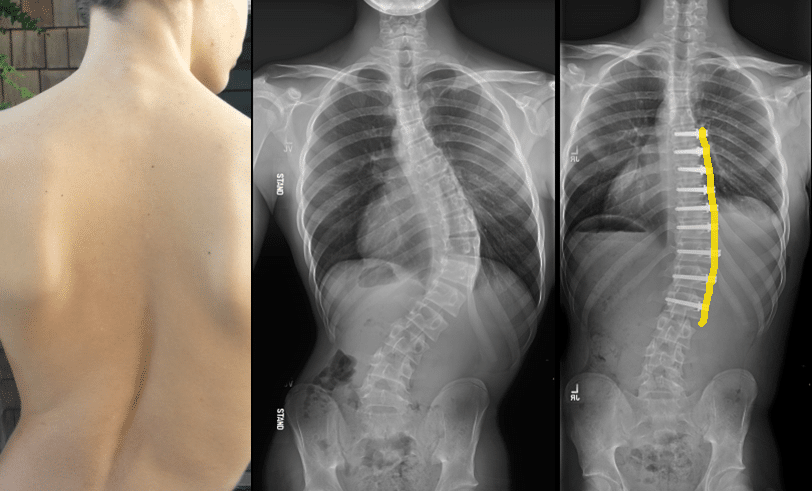
Severe Scoliosis X Ray:Severe scoliosis is a complex spinal condition characterized by a significant curvature of the spine. It can have a profound impact on a person’s quality of life, causing pain, discomfort, and functional limitations. In order to effectively diagnose and treat severe scoliosis, healthcare professionals rely on various imaging techniques, with X-rays playing a crucial role in the process. X-rays provide valuable information about the degree of curvature, structural abnormalities, and spinal rotation, which are essential for treatment planning. In this article, we will explore what severe scoliosis looks like on X-rays and its impact on treatment planning.

Understanding Severe Scoliosis
Scoliosis is a condition that causes an abnormal sideways curvature of the spine. While mild cases of scoliosis may not cause significant symptoms or require treatment, severe scoliosis can lead to pain, breathing difficulties, and even organ compression. Severe scoliosis is typically defined as a curvature of the spine greater than 40 degrees. It can occur in both children and adults, with different underlying causes and treatment approaches.
Importance of X-Rays in Diagnosis
Severe Scoliosis X Ray:X-rays are an essential tool in diagnosing severe scoliosis. They provide a detailed image of the spine, allowing healthcare professionals to accurately measure the degree of curvature and identify any structural abnormalities. X-rays also help in assessing spinal rotation and torsion, which can further impact treatment decisions. Without X-rays, it would be challenging to determine the severity of scoliosis and develop an appropriate treatment plan.

X-Ray Imaging Techniques for Severe Scoliosis
When it comes to imaging severe scoliosis, there are several X-ray techniques that healthcare professionals may use. The most common technique is the standing full-length X-ray, which captures the entire spine from the neck to the pelvis. This allows for a comprehensive evaluation of the curvature and alignment of the spine. In some cases, additional X-rays may be taken from different angles or in different positions to obtain a more detailed view of the spine.
Analyzing X-Rays: Key Findings
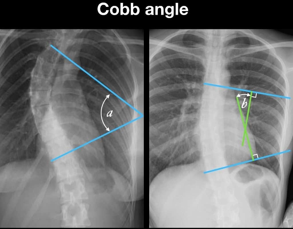
Severe Scoliosis X Ray: When analyzing X-rays of severe scoliosis, healthcare professionals look for several key findings that help guide treatment planning. The first and most important finding is the degree of curvature, measured in degrees using a method called Cobb angle measurement. This measurement helps determine the severity of scoliosis and whether treatment is necessary. Additionally, X-rays can reveal structural abnormalities such as wedged vertebrae, rotated vertebrae, or fused ribs, which can impact treatment decisions.
Visualizing Severe Scoliosis on X-Rays
Severe Scoliosis X Ray:Severe scoliosis is visually evident on X-rays, with the spine appearing curved rather than straight. The curvature can be either C-shaped or S-shaped, depending on the location and direction of the curve. The X-ray image allows healthcare professionals to visualize the entire spine and assess the extent of the curvature. This visual representation is crucial in understanding the severity of scoliosis and planning appropriate treatment.

Assessing the Degree of Curvature
Severe Scoliosis X Ray: One of the primary purposes of X-rays in severe scoliosis is to measure the degree of curvature. The Cobb angle measurement is used to determine the angle between the most tilted vertebrae at the top and bottom of the curve. This measurement helps classify scoliosis as mild, moderate, or severe, and guides treatment decisions. For example, a curvature of less than 25 degrees may not require treatment, while a curvature of more than 40 degrees often necessitates intervention.
Identifying Structural Abnormalities
X-rays also help identify structural abnormalities associated with severe scoliosis. These abnormalities can include wedged vertebrae, where the bones of the spine become triangular-shaped instead of rectangular, or rotated vertebrae, where the bones twist around their axis. Additionally, X-rays can reveal fused ribs, which can further contribute to the curvature of the spine. Identifying these structural abnormalities is crucial in determining the appropriate treatment approach.
Evaluating Spinal Rotation and Torsion
Severe scoliosis often involves not only a sideways curvature but also spinal rotation and torsion. X-rays allow healthcare professionals to evaluate the degree of rotation and torsion present in the spine. This information is important for treatment planning, as it can impact the choice of surgical techniques or the use of braces. By assessing spinal rotation and torsion, healthcare professionals can develop a more personalized and effective treatment plan for each individual.
Implications for Treatment Planning
The information obtained from X-rays of severe scoliosis has significant implications for treatment planning. Based on the degree of curvature, structural abnormalities, and spinal rotation, healthcare professionals can determine the most appropriate treatment approach. In mild cases, non-surgical interventions such as physical therapy, bracing, or exercise may be recommended. However, in severe cases, surgical intervention may be necessary to correct the curvature and prevent further progression.
Surgical Considerations for Severe Scoliosis
When severe scoliosis requires surgical intervention, X-rays play a crucial role in planning the procedure. X-rays help surgeons visualize the exact location and extent of the curvature, allowing them to determine the best approach for correction. They also aid in assessing the flexibility of the spine, which can influence the choice of surgical techniques. By analyzing X-rays, surgeons can develop a surgical plan that addresses the specific needs of each patient and maximizes the chances of a successful outcome.
Conclusion
Severe scoliosis is a complex spinal condition that requires careful diagnosis and treatment planning. X-rays are an invaluable tool in this process, providing essential information about the degree of curvature, structural abnormalities, and spinal rotation. By analyzing X-rays, healthcare professionals can accurately assess the severity of scoliosis and develop a personalized treatment plan. Whether it involves non-surgical interventions or surgical correction, X-rays play a crucial role in ensuring the best possible outcomes for individuals with severe scoliosis.
References
- National Scoliosis Foundation. “Understanding Severe Scoliosis and X-Ray Imaging.” Available at: https://www.scoliosis.org/severe-scoliosis-xray-imaging
- American Academy of Orthopaedic Surgeons. “Scoliosis: Diagnosis and Treatment.” Available at: https://www.aaos.org/Orthopedic-Conditions/Scoliosis
- Mayo Clinic. “Scoliosis: Diagnosis and Imaging Techniques.” Available at: https://www.mayoclinic.org/diseases-conditions/scoliosis/diagnosis-treatment/drc-20351054
- Spine-Health. “Severe Scoliosis and Its Imaging.” Available at: https://www.spine-health.com/conditions/scoliosis/severe-scoliosis-imaging
- Journal of Bone and Joint Surgery. “The Role of X-Rays in Scoliosis Management.” Available at: https://jbjs.org/content/role-x-rays-scoliosis
- RadiologyInfo.org. “Scoliosis X-Ray Examination: What to Expect.” Available at: https://www.radiologyinfo.org/en/info/scoliosis-xray
- Children’s Hospital of Philadelphia. “Imaging for Severe Scoliosis: Techniques and Findings.” Available at: https://www.chop.edu/conditions-diseases/severe-scoliosis-imaging
- Scoliosis Research Society. “X-Ray Analysis and Scoliosis Treatment Planning.” Available at: https://www.srs.org/professionals/xray-analysis-scoliosis
- British Journal of Radiology. “Diagnostic Imaging for Severe Scoliosis.” Available at: https://www.bjr.birjournals.org/content/diagnostic-imaging-severe-scoliosis
- Harvard Health Publishing. “How X-Rays Help in Managing Severe Scoliosis.” Available at: https://www.health.harvard.edu/x-rays-severe-scoliosis

