
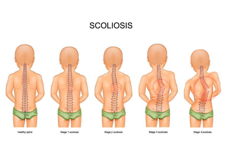
Scoliosis is a medical condition characterized by an abnormal curvature of the spine. It is typically categorized into three forms based on age of onset: infantile, juvenile, and adolescent idiopathic scoliosis. The latter, which affects children aged 10 to 18, is the most prevalent form [1].
The Importance of X-Ray Imaging in Severe Scoliosis Cases
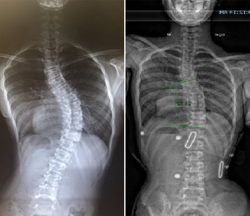
X-ray imaging is essential for diagnosing and managing severe scoliosis. It provides detailed visualization of spinal curvature, enabling accurate measurement of the Cobb angle and identification of structural abnormalities [2]. This imaging modality also assists in monitoring disease progression and evaluating treatment effectiveness [3].
Analyzing X-Ray Images: Key Measurements and Parameters
Key measurements from X-ray images include the Cobb angle, which quantifies spinal curvature. A Cobb angle greater than 40 degrees indicates severe scoliosis [4]. Other important parameters include the Risser sign, which assesses skeletal maturity, and vertebral rotation, which can influence treatment strategies [5].
Assessing the Severity of Scoliosis: Cobb Angle and Beyond
The Cobb angle is a standardized measurement for scoliosis severity, but additional factors like the Risser sign and vertebral rotation are also crucial. These measurements help in predicting disease progression and determining appropriate interventions [6][7].

Identifying Structural Abnormalities in Severe Scoliosis X-Rays
X-ray imaging can reveal structural abnormalities such as vertebral wedging and rib humping. Vertebral wedging involves triangular-shaped vertebrae, while rib humping results in uneven rib protrusion. Identifying these abnormalities is vital for treatment planning and surgical preparation [8][9].
Evaluating Spinal Curvature and Rotation in X-Ray Imaging
Severe scoliosis often involves both spinal curvature and rotation. X-ray imaging allows for the assessment of spinal rotation, which affects rib cage symmetry and lung function. Accurate evaluation is critical for effective treatment [10][11].
Examining the Impact of Severe Scoliosis on the Rib Cage
The impact of severe scoliosis on the rib cage includes asymmetry and deformities, such as rib fusion or rib flaring. X-ray imaging helps in understanding these changes and their implications for the spinal deformity and surrounding structures [12].
Assessing the Effects of Severe Scoliosis on Lung Function
Severe scoliosis can compromise lung function by reducing the space available for lung expansion. X-ray imaging provides insights into the degree of lung compression, aiding in the assessment of respiratory function and the need for potential interventions [13][14].
Interpreting X-Ray Findings: Implications for Treatment Options
The interpretation of X-ray findings is crucial for determining treatment options. Mild scoliosis may be managed with physical therapy and bracing, while severe cases often require surgical intervention. X-ray imaging helps in assessing the effectiveness of non-surgical treatments and guiding surgical decisions [15][16].

Surgical Intervention for Severe Scoliosis: X-Ray Guidance
X-ray imaging is pivotal in surgical planning and execution. Preoperative X-rays provide detailed information on spinal deformity, guiding surgical approach. Intraoperative X-rays are used to confirm correction and ensure accurate placement of implants [17][18].
Monitoring Progress and Treatment Success through X-Ray Imaging
Regular X-ray imaging is essential for monitoring scoliosis progression and evaluating treatment success. Follow-up X-rays help in assessing changes in spinal curvature, the effectiveness of interventions, and making necessary adjustments to the treatment plan [19][20].