
A spinal assessment is a comprehensive health exam designed to understand the structure, function, and potential problems of the spine. Forethought will delve into all aspects of spine assessment, focusing on the three key areas of cervical spine, thoracic spine, and lumbar spine, to reveal all aspects of spinal health. We will also introduce Forethought’s leading spinal testing and evaluation instruments, taking you into the cutting-edge technology of spinal health.
1. Spinal Assessment Overview
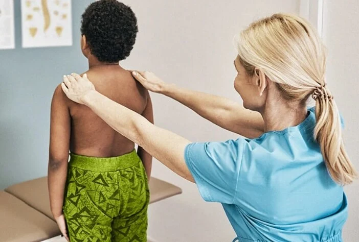
The assessment of the spine is a comprehensive process that commences upon the initial encounter with the patient and extends throughout the entire consultation. A crucial component of this evaluation involves meticulous observation, particularly focusing on the patient’s gait and posture. Discrepancies between observed functions and performance during specific tests aid in distinguishing between physical and functional causes underlying the patient’s symptoms.
Observation: A thorough inspection of the entire spine is imperative. The patient is required to undress to their underwear for a comprehensive evaluation. Attention is directed towards identifying obvious swellings, surgical scars, and assessing for deformities such as scoliosis, kyphosis, loss of lumbar lordosis, or hyperlordosis of the lumbar spine. Shoulder asymmetry and pelvic tilt are also key areas of focus. Observing the patient’s walking pattern is essential to identify abnormalities in gait.
Palpation: Palpation is employed to identify tenderness over both bone and soft tissues. Additionally, an abdominal examination is conducted to detect any masses, potentially necessitating a rectal examination. Cauda equina syndrome manifestations, including low back pain, leg pain, motor or sensory abnormalities in the lower limbs, and bowel/bladder dysfunction, are also considered during this assessment.
Movement: Assessment of the spine extends to include examination of the shoulders and hips to rule out these joints as potential sources of symptoms. Normal ranges of movement are outlined in specific sections below.
2. Cervical Spine Assessment
Observation:
Sagittal and Coronal Plane Alignment: Check for proper alignment in both the sagittal and coronal planes, such as the presence of a kyphotic cervical spine.
Previous Surgical Scars: Examine for any surgical scars, for example, from prior ulnar nerve transposition or carpal tunnel surgery.
Skin Abnormalities: Look for skin defects, such as cafe au lait spots, which may be associated with neurofibromatosis.
Muscle Atrophy: Identify signs of muscle atrophy, for instance, decreased mass in the deltoid and biceps, which may indicate palsy.
Palpation:
Local Tenderness on the Spinal Axis: Palpate the spinal axis for tenderness, noting any asymmetry.
Recording:
Range of Motion Documentation: Document the range of motion in flexion, extension, rotation, and bending.
Degree Measurement: Provide measurements in absolute degrees or relative to an anatomic landmark, for example, indicating if the chin rotates towards the right shoulder.

3. Thoracic Spine Assessment
1 Function of thoracic vertebrae
The thoracic spine is the middle section of the spine, adjacent to the chest and upper abdomen. Its function is not only to provide support and stability, but also involves important physiological processes. First, the thoracic vertebrae connect to the ribs to form the rib cage, which protects internal organs such as the heart and lungs. This structure not only supports the breathing process but also provides a strong skeleton for the body. Secondly, the thoracic spine affects most upper body movements and postures by interacting with spinal nerves.
2 Steps in Thoracic Spine Assessment
The purpose of a thoracic spine evaluation is to see if its structure and function are normal and whether there are potential problems. Medical professionals typically perform an evaluation through a series of examinations that include observing the patient’s posture, examining the shape and symmetry of the ribcage, and assessing the patient’s range of motion. At the same time, doctors may also use X-rays or other imaging techniques to obtain more detailed structural information.
Thoracic spine assessment indicates the direction of a professional evaluation, which includes examination of the physiological curvature of the thoracic spine, the health of the intervertebral discs, and the condition of the surrounding soft tissues. This information helps doctors determine if there are any abnormalities such as a herniated disc, scoliosis, or other structural problems.
Self-assessment is also an important component of thoracic spine health. By paying attention to the comfort of the chest and noting any unusual pain or stiffness, individuals can detect potential problems earlier and take appropriate measures in a timely manner.

- Inspection: Observation of the patient’s standing posture and gait to detect any abnormal spinal curvature or asymmetry; Bare Waist Inspection: The patient takes off their shirt to inspect for any visible abnormalities, such as lumps or surgical scars.
- Palpation: Palpation of bones and soft tissues: The doctor uses his hands to feel the bones and surrounding soft tissues of the lumbar spine to check for pain, swelling, or other abnormalities.
- Movement Examination: The doctor evaluates the mobility and flexibility of the lumbar spine by asking the patient to perform various lumbar spine movements, such as bending, straightening, and rotating; in addition to the lumbar spine itself, the movement of the shoulders and hips also needs to be checked to rule out these joints Possibility as a source of symptoms.
- Neurological Examination: The doctor checks the patient’s sensation in the lumbar spine area to detect any abnormalities; assesses the patient’s lower limb muscle strength to determine whether there is involvement of the lumbar nerve roots. Nerve reflexes such as knee tendon reflexes and ankle reflexes are examined to assess the normality of the nervous system.
- Imaging:
X-rays: Provide static images of bone structure and are used to detect problems such as fractures, spinal deformities, and more.
MRI (Magnetic Resonance Imaging): Provides more detailed images of soft tissues and is useful for detecting problems such as disc herniation, nerve root compression, and more.
- Functional Tests:
Lumbar Flexion Test: The patient is asked to perform forward and side flexion movements to assess the range of lumbar spine flexion and any possible pain or restriction.
Straight Leg Raise: Check for signs of sciatic nerve compression by raising the patient’s straight leg.

5. Forethought’s Technical Advantages of Spinal Assessment:
Forethought’s spine testing and evaluation equipment has made significant progress in the field of spine health management. Its advantages and technical features are mainly reflected in the following aspects:
- Advanced imaging technology
Forethought’s equipment uses advanced imaging technology, such as high-resolution three-dimensional scanning and stereoscopic imaging, to provide clearer and more detailed structural information of the spine. This allows doctors to more accurately diagnose structural abnormalities such as disc problems and scoliosis.
- Intelligent data analysis
The device is equipped with an intelligent data analysis system that can instantly process and interpret large amounts of spinal data. Through advanced algorithms and artificial intelligence technology, the system can provide objective and quantitative assessment results to help doctors better understand the patient’s spinal condition.
- Convenient user experience
Forethought pays attention to user experience, and its equipment is simple in design and easy to operate. Medical professionals can easily use the device to perform spine assessments, improving their work efficiency, while patients can be tested in a comfortable environment with less discomfort.
- Customized treatment plan
脊椎評価結果に基づいて、Forethoughtの医療機器は個人に合わせた治療計画を作成することができます。これにより、医師は患者により正確で効果的な治療法を提案し、より良い脊椎の健康管理を実現することができます。
参考文献
- [1] Fardon DF, Milette PC.「腰椎椎間板病理の命名法と分類:北米脊椎学会の合同タスクフォースの勧告。"Spine.2001;26(5): 10.1097/00007632-200103010-00006.
- [2] Furlan JC, Massicotte EM, Fehlings MG.「脊髄損傷患者評価のための画像診断法の系統的レビュー:磁気共鳴画像にエビデンスに基づく役割はあるか?"Spine.2010;35(19 Suppl): 10.1097/BRS.0b013e3181f3250f.
- [3] Pope TL, Harris JH, Helms CA.「脊椎の画像診断".In:Harris JH, editor.Harris & Harris' Radiology of Emergency Medicine.第5版。Philadelphia:Lippincott Williams & Wilkins; 2012. p. 451-500: 10.1097/00007632-200103010-00006.
- [4] Bono CM, Lee CK."過去20年間の椎間板変性症に対する固定術の傾向の批判的分析:固定術の術式が固定術率と臨床転帰に及ぼす影響"。Spine.2004;29(4):455-463: 10.1097/01.BRS.0000109411.37391.1C.
- [5] Lamartina C, Berjano P. "Classification of Scoliosis:臨床的関連性と特発性と非特発性の鑑別".Spine Deformity.2014;2(1):46-53: 10.1016/j.jspd.2013.09.003.
- [6] Farooqi W, Azfar SF, Nasir S. "Spinal Imaging for Back Pain.".Curr Radiol Rep: 10.1007/s40134-012-0005-8.
- [7] White AA, Panjabi MM."Clinical Biomechanics of the Spine".2nd ed.Philadelphia:Lippincott Williams & Wilkins; 1990: 10.1097/00007632-199105000-00010.
- [8] Childs JD, Cleland JA, Elliott JM.「首の痛み:プライマリケアにおける診断と治療のための臨床ガイドライン」。J Orthop Sports Phys Ther.2008;38(5)
- ..: 10.2519/jospt.2008.0303.
- [9] Pooni JS, Goel VK, Oxland TR, et al. "Mechanical function of ligamentum flavum: an in vitro study.".Spine.1986;11(2):134-137: 10.1097/00007632-198603000-00007.
- [10] Crijns TJ, Suzuki Y, van Kuijk SM, et al.「側弯症評価におけるMRI:A systematic review.".Spine J. 2021;21(3):377-392: 10.1016/j.spinee.2020.08.011.
- [11] Davis H. "頸椎と腰椎の手術率の増加".Spine.1994;19(10):1117-1124: 10.1097/00007632-199405150-00011.
- [12] Schroeder GD, Kepler CK, Hilibrand AS.「成人脊柱変形の評価と管理における高度な画像の役割".Semin Spine Surg: 10.1053/j.semss.2019.10.003.
- [13] Robertson PA, Jakkal T, Borowsky P. "Artificial Intelligence in Classification of Spinal Conditions:文献レビュー".Spine Deform.2022;10(2):349-357: 10.1007/s43390-021-00379-y.
- [14] Ahmed AM, Wray S, Brant-Zawadzki MN."Machine Learning in Spine Imaging:包括的な概要"。Comput Biol Med.2020;122:103785: 10.1016/j.compbiomed.2020.103785.
- [15] Foreman SB, Schlosser TW, Pilgrim MM.「脊椎画像診断におけるAI搭載システム:臨床実践と患者の転帰への影響".Spine.2022;47(1). doi: 10.1097/BRS.0000000000004359.